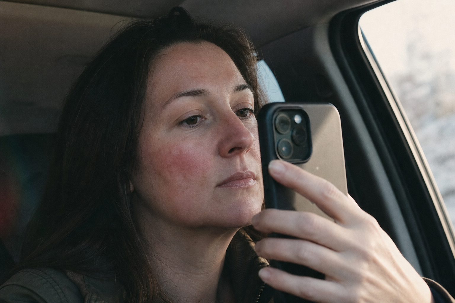
The two weeks before your rosacea appointment are for photos, not food diaries.
A GP said 'could be rosacea, come back in two weeks.' The log that helps most is timestamped photos plus context, built to show phenotype, not a two-month food diary.
Two weeks, and a blank notes app
A GP glances at your cheeks for maybe ninety seconds, says the flushing across your nose and cheeks could be rosacea, and tells you to come back in two weeks with a sense of the pattern. Then you're in the car park with your phone open, already typing what you had for lunch.
That instinct, log the food, is the wrong first move. And it's the one nearly every rosacea guide online will hand you. The stretch between a suspected-rosacea comment and a first dermatology appointment is the single highest-value documentation window in the whole timeline of this condition. Most people spend it building the wrong file.
The pre-diagnosis window is a phenotype problem, not a trigger problem
Call it the pre-diagnosis log. At a first appointment, the dermatologist isn't trying to work out what sets your skin off. They're answering one question: is this rosacea, and if so, what does it actually look like on your face. That's phenotype identification. Phenotype just means the set of features your skin shows and that you feel, the redness, the bumps, the visible vessels, the eye irritation, as opposed to sorting you into a named subtype.
Trigger elimination comes later, after a diagnosis exists. So the log that earns its place in these two weeks is one built to show phenotype: timestamped photos across fourteen days, paired with the short context that explains what a camera can't. Not two months of a food diary that answers a question nobody's asking yet.
Why the 'keep a rosacea diary' advice misfires here
The entire keep-a-diary content universe is written for people who already have a diagnosis and are managing a known condition. Food-and-flush logs, red-wine-versus-white debates, elimination charts. Useful, later.
For the pre-diagnosis reader it's the wrong tool twice over. It optimizes for trigger elimination when the appointment is about identification, and it takes too long to pay off, two months of accumulation for a two-week window. It also drops the one thing a clinician most needs. 'Cheeks were red on Tuesday' is a sentence. A timestamped photo of your cheeks on Tuesday is evidence.
What a dermatologist is actually reading in fifteen minutes
In 2017 the field changed how rosacea gets diagnosed. The National Rosacea Society expert committee (Gallo et al., JAAD 2018) and the global ROSCO panel (Tan et al., Br J Dermatol 2017) both moved away from sorting patients into four numbered subtypes and toward a phenotype approach: you diagnose from the features that are present, not from a bucket you get assigned.
Two features are diagnostic on their own. Persistent centrofacial erythema, meaning lasting redness in the middle of the face, and phymatous changes, meaning skin thickening, most often on the nose. Several others count as major features that support the picture: transient flushing, papules and pustules, telangiectasia (visible small blood vessels), and ocular involvement (eye symptoms). A first appointment is short, often around fifteen minutes, much of it history and looking. Fourteen days of photos do the looking for the days the clinician wasn't in the room.
Feature
2017 role in diagnosis
What it looks like
Persistent centrofacial erythema
Diagnostic on its own
Lasting redness across the central face that doesn't fully settle
Phymatous changes
Diagnostic on its own
Thickened skin, most often on the nose
Flushing / transient erythema
Major feature
Redness that comes and goes, often with heat or emotion
Papules and pustules
Major feature
Small bumps and pus-filled spots, no blackheads
Telangiectasia
Major feature
Visible small blood vessels near the surface
Ocular involvement
Major feature
Dry, gritty, irritated or bloodshot eyes
The 2017 phenotype approach. Source: Gallo et al., JAAD 2018; Tan et al. (ROSCO), Br J Dermatol 2017.
Redness reads differently on darker skin
On Fitzpatrick IV to VI skin, rosacea's redness can look dusky or violaceous rather than pink, and it's under-recognized as a result. Adamson & Smith (JAMA Dermatology 2018) and Daneshjou et al. (2022) documented that dermatology image algorithms, trained mostly on lighter skin, fail harder on darker skin. That's why what you feel, your sensory phenotype (burning, stinging, dryness, gritty eyes), belongs in the log right next to the photo.
What to capture, per flare, over fourteen days
Here's the shape of it in practice. Each time your face flares or simply looks different from baseline, spend two minutes: take a photo in consistent, natural light, let the timestamp record itself, and add four short context notes. Do that whenever it happens, not on a schedule.
The context is what turns a picture into a readable record. If you catch the same central redness at 8am and 8pm across several days, that pattern reads as persistent centrofacial erythema, one of the diagnostic features. If the redness only shows up after a hot shower or a workout and fades, that's flushing, a different major feature. The photo alone can't tell those two apart. The photo plus the timestamp and the heat note can.
Capture per flare
What it tells the clinician
Photo in consistent, natural light
Where redness sits and whether it's persistent or transient
Timestamp (automatic)
Time-of-day pattern across the fourteen days
Ambient temperature / heat exposure
Separates heat-driven flushing from lasting erythema
Products applied that day
Flags an irritant reaction a derm can rule in or out
Sleep hours and stress, as numbers
Turns a vague 'bad week' into a trend that can be read
A per-flare capture set for the pre-diagnosis window.
Fourteen good days beat two vague months
If this holds, the reader stops treating the pre-diagnosis window as a trigger hunt and starts treating it as evidence-gathering for phenotype. The payoff is concrete. The dermatologist reaches a diagnosis faster and with more confidence, and you walk out with a named phenotype and a starting plan, rather than being told to go track for another two months.
One feature is worth logging on purpose, because it's the one most often missed: ocular involvement. Dry, gritty, or irritated eyes get written off as unrelated, and yet the 2017 criteria list them as a major feature. If your eyes have been bothering you alongside the flushing, capture that too. It may be the detail that completes the picture.
Where Skinframe fits
We built Skinframe around this exact window. It timestamps every photo on your device, photos stay on your phone, keeps the framing and light consistent, and pairs each shot with the short context fields that turn a picture into something a clinician can read in a first visit.
We don't ship a face-scanning skin analysis, and the equity literature is the reason. Adamson & Smith (JAMA Dermatology 2018) and Daneshjou et al. (2022) both documented that dermatology image algorithms fail harder on darker skin because of what they were trained on. Rosacea is already under-recognized on Fitzpatrick IV to VI skin, so we lean on your own sensory phenotype, what you feel, next to the photos, instead of a machine guessing from a picture. None of this is a diagnosis, and it isn't meant to be. It's the file you hand across the desk. Bring it, and let your dermatologist read it.
Skinframe is built for the two weeks before your appointment: timestamped photos, on-device, paired with the context a dermatologist can read. Start your fourteen-day log.
We read the 2017 rosacea reclassification (Gallo et al., JAAD 2018; Tan et al., ROSCO 2017) so the log we built answers the question a first appointment actually asks: what does this look like, not what sets it off. Photos stay on your phone, timestamped, ready to hand across the desk.