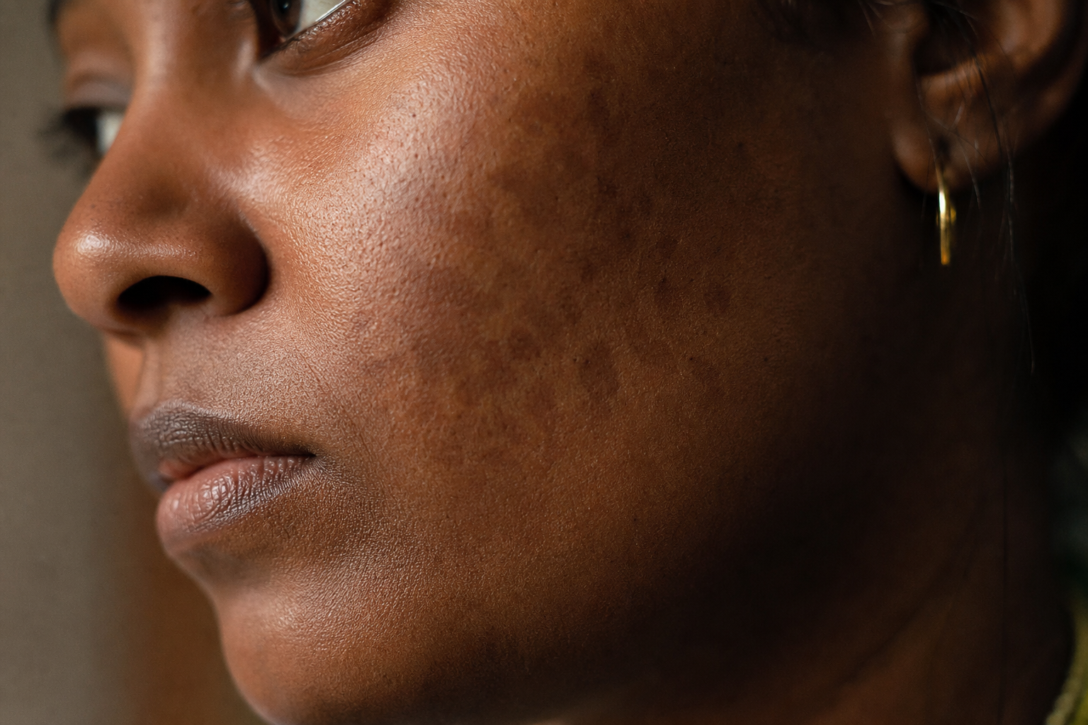
The mark left after a rosacea flare isn't always active rosacea
In medium and dark skin, post-inflammatory pigment can outlast a rosacea flare by weeks. Without dated photos, a single exam can't tell remission from relapse.
Six weeks out, the cheek still holds color
Six weeks after the flare, the burning is gone and the papules have flattened, but the cheeks still hold color: flat, brown-toned patches where the worst of the inflammation sat. The dermatology note from the last visit reads *still active, let's escalate*, and a new prescription follows. On deeper skin, that reading is often wrong. The color left behind is post-inflammatory pigment, flat discoloration the skin lays down while it heals, long after the active flare has settled.
Call it the aftermark
When inflammation settles, skin with more melanin frequently responds by depositing extra pigment at the site: a flat brown or gray-brown discoloration dermatologists call post-inflammatory hyperpigmentation, or PIH. It sits exactly where the flare was hottest. It can linger for weeks after every papule and every trace of stinging is gone, and on medium and deep skin it reads, at a glance, a lot like the redness that caused it. On those complexions, that window of persistence often stretches longer than patients expect, sometimes well beyond what lighter skin would show for the same flare. The hardest thing about the aftermark is timing. A flare and its pigment aftermath look similar in a mirror, and a single point-in-time exam captures only one frame of a process that plays out over months.
Why 'more color means more disease' breaks on deeper skin
The dominant framing treats visible color on the face as a proxy for disease activity. More color, more rosacea, more treatment. That shortcut mostly holds on fair skin, where post-inflammatory pigment is faint and fades fast, so residual color really does track with active flushing. It breaks on deeper skin. Here the pigment response is stronger and slower to clear, and it can outlast the inflammation by a wide margin. A clinician working from one appointment, or a patient working from one glance in the mirror, sees color and reasonably reads it as ongoing rosacea. So the treatment escalates on skin that has already reached remission, and the patient carries the side effects of a therapy aimed at a flare that ended weeks ago.
What the literature and the search data actually say
Rosacea in skin of color has a documented recognition problem. Maliyar and colleagues (2022) reviewed how rosacea presents across Fitzpatrick IV to VI and found the classic redness cue that anchors most diagnostic thinking is muted against more melanin, which pushes the condition toward under-recognition and mistiming. The same melanin that dampens the redness signal amplifies the pigment sequel. That's the trap the search data keeps surfacing: one of the most common People-Also-Ask queries in this space is *what is commonly mistaken for rosacea*, and post-inflammatory pigment is a recurring answer for darker complexions.
There's a second reason we don't lean on an automated read of a face photo to settle this. Adamson and Smith (JAMA Dermatology, 2018) documented that dermatology algorithms trained largely on light skin fail measurably harder on dark skin, and Daneshjou and colleagues (2022) showed how thin the diverse-image training data actually is. A tool that claims to grade redness from a photo inherits exactly those blind spots, on exactly the skin tones where the redness-versus-pigment distinction matters most. The dependable instrument here is a dated record a clinician can interpret, and the photo's value is as evidence, not as an automated grade.
The one test you can run and document
There's one test a patient can run at home and document, and it's the cleanest way to tell the two apart: gentle pressure. Press a clean fingertip or a clear glass slide against the patch for a second or two, then look. Active erythema, the redness of a live flare, blanches, meaning the color briefly whitens because you're pressing blood out of the dilated vessels underneath. Post-inflammatory pigment doesn't blanch, because it's melanin sitting in the skin rather than blood in a vessel. Dermatologists call this maneuver diascopy. It's a signal worth capturing, not a diagnosis.
Run it as a sequence, not a one-off. A photo at flare onset, one at the peak, one when the stinging stops, and one a few weeks later, all dated, turns a guess into a timeline. If week-eight color blanches, there's likely still vascular activity. If it doesn't, the flare has resolved and what remains is the aftermark healing on its own schedule.
Sign
Active erythema
Post-inflammatory pigment
Response to gentle pressure (diascopy)
Blanches, whitens briefly
Does not blanch
Underlying cause
Dilated blood vessels
Melanin deposited in the skin
Typical timing
During an active flare
After the flare settles
What it signals
Disease activity
Healed sequel of past inflammation
How active erythema and post-inflammatory pigment differ. General dermatology; confirm with a clinician.
The blanch test documents, it doesn't diagnose
Diascopy is a way to capture a signal, not to rule rosacea in or out on your own. Use it to build the record you bring to your dermatologist, and don't start, stop, or escalate any treatment based on it alone.
Without a dated timeline, both patient and dermatologist are guessing at which phase the skin is actually in.
What changes if the aftermark is real
If the aftermark is real and common, the whole picture of what a rosacea visit needs changes. The most useful thing a patient can bring to a deeper-skinned rosacea appointment is a record of how the skin changed over time, more than a description of how it looks on the day. A dated sequence lets a dermatologist separate disease from its sequel, hold treatment when the skin is actually in remission, and address lingering pigment as pigment, with the tools that target melanin, rather than escalating a rosacea therapy against color that no anti-inflammatory will touch.
A timeline a single exam can't reconstruct
This is the specific gap we built Skinframe to close. The app is a dated, on-device photo log: you capture the same area under consistent framing, and every image carries its own timestamp, so onset, peak, and resolution line up as a sequence a dermatologist can scroll through. We deliberately don't grade your redness or tell you what phase you're in, for the reasons Adamson and Smith and Daneshjou spelled out. The photos stay on your device. The judgment stays with your clinician. What Skinframe adds is the one thing a single exam can't reconstruct after the fact: the timeline that shows whether the color on your cheek today is the flare or its shadow.
What we're watching next
We're watching the consensus literature for a skin-of-color-specific update to rosacea grading. The 2017 phenotype framework moved the field away from subtypes, but it still leans on erythema as the anchor feature, and the pigment-sequel problem in Fitzpatrick IV to VI skin sits mostly outside it. If a society statement formalizes how to score the aftermark separately from active disease, this is the first article we'll revise. None of this replaces an exam. The blanch test and the photo log are there to make the conversation with your dermatologist more precise, not to substitute for it, so bring your record and talk to your dermatologist.
Start a dated photo record now, so your next dermatologist visit reads from a timeline instead of a single snapshot.
Skinframe was built with the rosacea skin-of-color literature open on the desk, from Maliyar 2022 on presentation across Fitzpatrick IV-VI to Adamson and Smith 2018 on why we refuse to auto-grade a face photo. It's a documentation tool for you and your dermatologist, not a diagnosis.