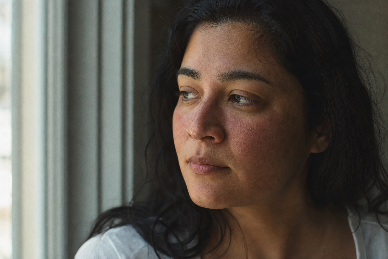
Rosacea has no lab test, so your photo log is the test.
Rosacea has no confirmatory test. A dermatologist reads it from visible phenotype features, which means a timestamped photo series is the only objective record you can actually hand them.
The exam room runs on what can be seen, not what was felt
Fifteen minutes. That is roughly the window a dermatologist has to decide whether the redness on your face is rosacea, and a written note that says "my cheeks were on fire Tuesday night" does almost nothing to fill it.
Rosacea has no blood panel, no swab, no biopsy marker that comes back positive. It is diagnosed entirely by looking. That single fact reshapes what you should be bringing to the appointment. Most people bring a story about how their skin has been behaving. The clinician cannot examine a story.
What they can examine is a record of what the skin actually looked like across time, in the moments you were not sitting in their chair. That record is a photograph, or better, a series of them.
The photo is the test
Here is the principle the rest of this follows from: when a condition has no confirmatory lab result, the objective visual record becomes the closest thing to one. Rosacea is that condition. Its diagnosis is pattern recognition applied to specific, nameable features on the face, and the only way those features travel from your bad flare night into the clinician's assessment is as an image.
Call it the photo-is-the-test principle. Not the photo as a nice supplement to the appointment. The photo as the primary evidence the appointment is structurally missing, because a flare that peaked at 9pm on a Tuesday is invisible to a Thursday-morning exam under office lights.
"How do you describe rosacea?" is the wrong question
Search for how to describe rosacea at a physical exam and you will find people trying to assemble the right words: blotchy, warm, stinging, worse after wine. The instinct is that sharper vocabulary will close the gap. It will not, because prose is the wrong medium for this job.
Language compresses. "Really red" flattens a specific intensity, location, and border pattern into two words, and it strips out the one axis rosacea diagnosis depends on most, which is time. A photograph preserves all of it at once: exactly where the erythema sits, how far it spreads, whether it fades or fixes. A dermatologist does not need your adjectives translated. They need the thing your adjectives were describing.
A story tells a dermatologist what you felt. A photo shows them what they have to diagnose.
What the diagnosis actually maps to
The 2017 update to the standard rosacea classification (Gallo et al., J Am Acad Dermatol 2018, the National Rosacea Society Expert Committee) sorted rosacea's presentation into features rather than the older subtype boxes. Two features are independently diagnostic on their own: fixed centrofacial erythema, meaning persistent redness in the center of the face, and phymatous changes, meaning thickening of the skin. Several others are major features that build the picture: papules and pustules, flushing (the transient kind), telangiectasia (visible dilated vessels), and ocular involvement.
Every one of those is a visual observation with a location, an intensity, and often a duration. That is precisely the data a timestamped photo captures and a sentence discards. The table below is the translation layer between the classification and what your camera is actually recording.
Location, baseline intensity, whether it stays between flares
Phymatous changes (skin thickening)
Diagnostic on its own
Contour and texture change over months
Papules and pustules
Major feature
Count and centrofacial distribution pattern
Flushing (transient redness)
Major feature
Onset and duration, only if the series is timestamped
Telangiectasia (dilated vessels)
Major feature
Which vessels are visibly involved and where
Ocular involvement
Major feature
Lid-margin and eye-area changes often missed verbally
Features per Gallo et al., J Am Acad Dermatol 2018 (2017 NRS classification). Right column is what an image preserves that prose does not.
Why the lighting is part of the evidence
A photo only works as evidence if it is honest about color, and that is where most home documentation quietly fails. Fluorescent office and bathroom lighting shifts and flattens red tones, which suppresses the exact erythema signal a clinician is trying to read. Consistent natural light, same window, same time of day, is not an aesthetic preference. It is what keeps one flare comparable to the next.
This matters most for darker skin. In Fitzpatrick IV to VI tones, rosacea erythema often reads as a dusky or violaceous discoloration rather than bright red (In skin of color, persistent facial erythema is reported less frequently than papules and pustules in rosacea patients. ([source](https://www.jaad.org/article/S0190-9622(18)32576-3/fulltext))), which is one reason rosacea is underrecognized in these patients. A consistent, well-lit series is doing more diagnostic work there, not less. And it should be a series a clinician reads, not a scan an algorithm judges: Adamson & Smith (JAMA Dermatol 2018) and Daneshjou et al. (Science Advances 2022) both documented that dermatology algorithms perform measurably worse on darker skin. The record we are describing is human-read evidence, deliberately.
Lighting is a diagnostic variable, not a filter
Fluorescent light suppresses red signal. Shoot in the same natural light each time (one window, similar time of day), no filters, no beauty modes. In darker skin, where erythema can present dusky or violaceous, inconsistent lighting can erase the finding entirely.
One appointment versus a flare-cycle series
Picture two versions of the same patient. The first walks in and says the redness comes and goes, worse some weeks, and shows the clinician a face that happens to be calm that morning. The exam sees a baseline and has to take the flaring on faith.
The second brings twelve photos across six weeks, same window light, each timestamped. Now the pattern is legible: centrofacial redness that persists at a low level and spikes on specific days, papules appearing in a consistent distribution, a flush that arrives and fades within an hour. That is pattern data across multiple flare cycles, and it is something a single fifteen-minute visit cannot generate no matter how skilled the clinician is. The appointment was never designed to observe you over time. The photo series is.
Build the record before you need it
If the diagnosis is a visual pattern read across time, the useful thing to own is a visual record kept across time. That is the whole design premise behind Skinframe. We built it to capture the phenotype features a dermatologist actually classifies against, in consistent light, timestamped, stored on your device rather than sent off to be scored by anything. No skin-tone-biased face scan, for the reasons Adamson & Smith and Daneshjou spelled out. Just a clean, comparable series you can hand across the desk.
A photo log will not tell you whether you have rosacea, and neither will we. It gives your dermatologist the objective evidence the condition otherwise leaves them without. The next step is the same one it always is with skin: bring the record, and talk to your dermatologist.
Start a timestamped photo record before your next flare, and walk into your appointment with evidence a dermatologist can actually read instead of a description they have to imagine.
Rosacea's diagnosis is defined by visible phenotype features (Gallo et al. 2018), and it has no confirmatory lab test. That is the entire case for treating your own photo series as evidence rather than a keepsake. Skinframe is built to capture exactly those features, in consistent light, timestamped, and kept on your device.