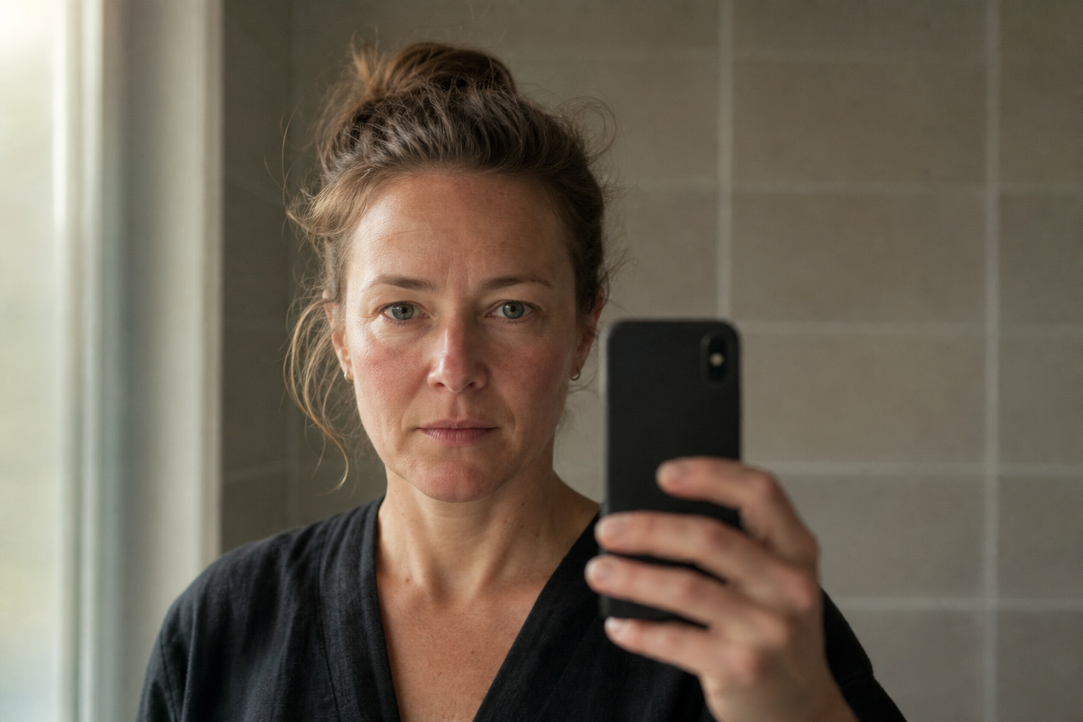
Make your rosacea flares readable in the ninety seconds your dermatologist has.
Rosacea has no lab test, so the diagnosis comes from your photo record. Three decisions turn a chaotic camera roll into a log a dermatologist can classify in one visit.
The flare is gone by the time the appointment starts
Your worst flare hit on a Tuesday three weeks ago. Cheeks hot, a rope of visible vessels along the nose, two papules you would have called acne if the burning had not told you otherwise. By the morning of your dermatology appointment, the skin is calm. Even, quiet, almost smug about it. You sit down for a ten-minute visit and try to describe something that is no longer on your face. This is the structural mismatch at the center of rosacea care: the condition lives across weeks of unpredictable flares, and the appointment is a short window on a day the skin usually behaves.
Rosacea is diagnosed from a record, not a moment
Here is what makes this condition different from a rash you can point to. Rosacea has no blood test, no swab, no biopsy that settles it. The 2017 reclassification from the National Rosacea Society (Gallo et al., JAAD 2018) moved diagnosis onto phenotype, the visible and felt features of the condition that replaced the older subtype labels. A dermatologist looks for persistent centrofacial redness that periodically intensifies, or phymatous skin changes, plus supporting features like flushing, papules, visible vessels, or eye involvement. Every one of those is a pattern over time, not a snapshot. So the evidence has to come from a photo record organized well enough for a dermatologist to classify in one visit. We call the target the ninety-second read: a log a clinician can scan in a minute and a half and come away knowing your phenotype. Most patients arrive with the opposite. Nothing, or a camera roll of four thousand images with no way in.
The problem is rarely too few photos
The standard advice is take pictures of your flares. It is not wrong, it is just the easy half. Volume is not evidence. A folder of forty flare photos shot at different distances, in different light, at different angles, with no note about what happened that week, is not a record a dermatologist can read. It is forty separate puzzles. The clinician cannot tell whether the redness got worse or the lighting did, whether the papule is new or the same one from Tuesday, whether the trigger was the wine or the sun or the stress or all three. The three weeks you spent capturing do not translate, because capture was never the hard part. Organizing what you captured is.
Three decisions that turn photos into a record
Three organizational choices separate a usable log from a pile of images, and you make them before the next flare, not after.
First, fix the frame. Same angle, same distance, same light source, every session. Front and both three-quarter profiles is enough. Consistency is what lets a dermatologist compare this Tuesday to the last one instead of guessing whether your camera moved. It matters most on deeper skin tones, where erythema (redness) photographs faintly and inconsistent lighting can wash out the one feature that carries the diagnosis. Rosacea might be underreported and underdiagnosed in skin of color populations due to difficulty discerning erythema and telangiectasia. ([source](https://www.jaad.org/article/S0190-9622(18)32576-3/fulltext)).
Second, log the metadata before the flare fades, not from memory two weeks later. What you ate and drank, heat and sun exposure, stress, where you are in your cycle if that tracks for you. Reconstructed triggers are guesses; triggers logged the same day are data. The 2017 framework counts burning and stinging as real features of rosacea, so note the sensory phenotype, what the skin feels like and not only how it looks, even on the days the camera catches nothing.
Third, compress. A dermatologist is not going to swipe through forty images. The record that works is a short timeline: a handful of representative flare days in order, each with its photo and one line of context, ending on today. That is the ninety-second read.
Decision
What noise looks like
What a usable log looks like
Frame
Different angle, distance, and lighting each time
Same angle, distance, light every session; front plus two profiles
Metadata
Triggers reconstructed from memory weeks later
Diet, heat, sun, stress, cycle logged the same day as the flare
Timeline
Four thousand photos, no order
A dozen representative days in sequence, one line of context each
The three decisions that separate a scannable rosacea log from an unnavigable camera roll.
Three weeks, one appointment, walked through
Say your appointment is a Thursday, three weeks out. Day one is calm skin, and you take the baseline anyway: front and both profiles, in the same bathroom light you will use every time. Friday night a warm room and a glass of wine bring a flush, so you photograph it that evening and note both. The following Wednesday brings papules and a burning you can feel from across the room. Photo, note, done in under a minute. You are not documenting every day. You are catching the four or five days that actually move, and shooting them the same way each time.
The morning of the appointment your skin is, of course, clear. It does not matter. You hand over a timeline: baseline, three flare days in order, each with its trigger line, ending on this quiet face. In ninety seconds the dermatologist sees persistent centrofacial erythema that intensifies with heat and alcohol, inflammatory papules, and the burning you reported that the camera never caught. That is a phenotype. That is a visit that ends in a plan instead of come back when it flares.
What a classifiable record changes
When the record is readable, the appointment stops being a memory test. You are not competing with your own clear skin or narrating a flare you can no longer see. The dermatologist classifies from evidence, which is the entire point of the phenotype approach, and the conversation moves to treatment on the first visit instead of the second or third. It changes what happens afterward too. The same timeline that got you classified becomes the baseline you measure treatment against, so the next appointment asks a sharper question: are the papules responding, not how have things been in general.
Where a tool earns its place
None of this requires an app. A consistent corner of the bathroom, a notes file, and the discipline to shoot before the flare fades will get you there. The friction is that the discipline is exactly what a flare week erodes, and reconstruction after the fact is where most logs fall apart. We built Skinframe around these three decisions so the structure holds when you do not feel like holding it: a fixed capture frame that keeps angle and light consistent shot to shot, trigger metadata attached to each photo the moment you take it rather than reassembled later, and a timeline that compresses weeks into the sequence a dermatologist can actually read. The photos stay on your device. We do not run skin analysis or guess your diagnosis; that is the dermatologist's job, and the log exists to give them something worth classifying.
Before your next appointment
Start with your next flare, not your appointment week. The record that reads in ninety seconds is built one consistent photo at a time, across the weeks when your skin is doing the talking. Bring it in, and let your dermatologist classify from evidence instead of from memory. And whatever the timeline shows, the diagnosis and the treatment plan are theirs to make: talk to your dermatologist.
Keep every flare on one timeline, angle and light consistent, triggers noted the day they happen, ready to hand your dermatologist. Start with your next flare.
Rosacea diagnosis is phenotype-based with no confirming lab test (Gallo et al., JAAD 2018), which puts the burden of evidence on the patient's own record. Skinframe is built around the three decisions that make that record classifiable: consistent framing, trigger metadata captured at shoot time, and a compressed timeline. Photos stay on your device, and there is no skin-analysis layer pretending to do the dermatologist's job.