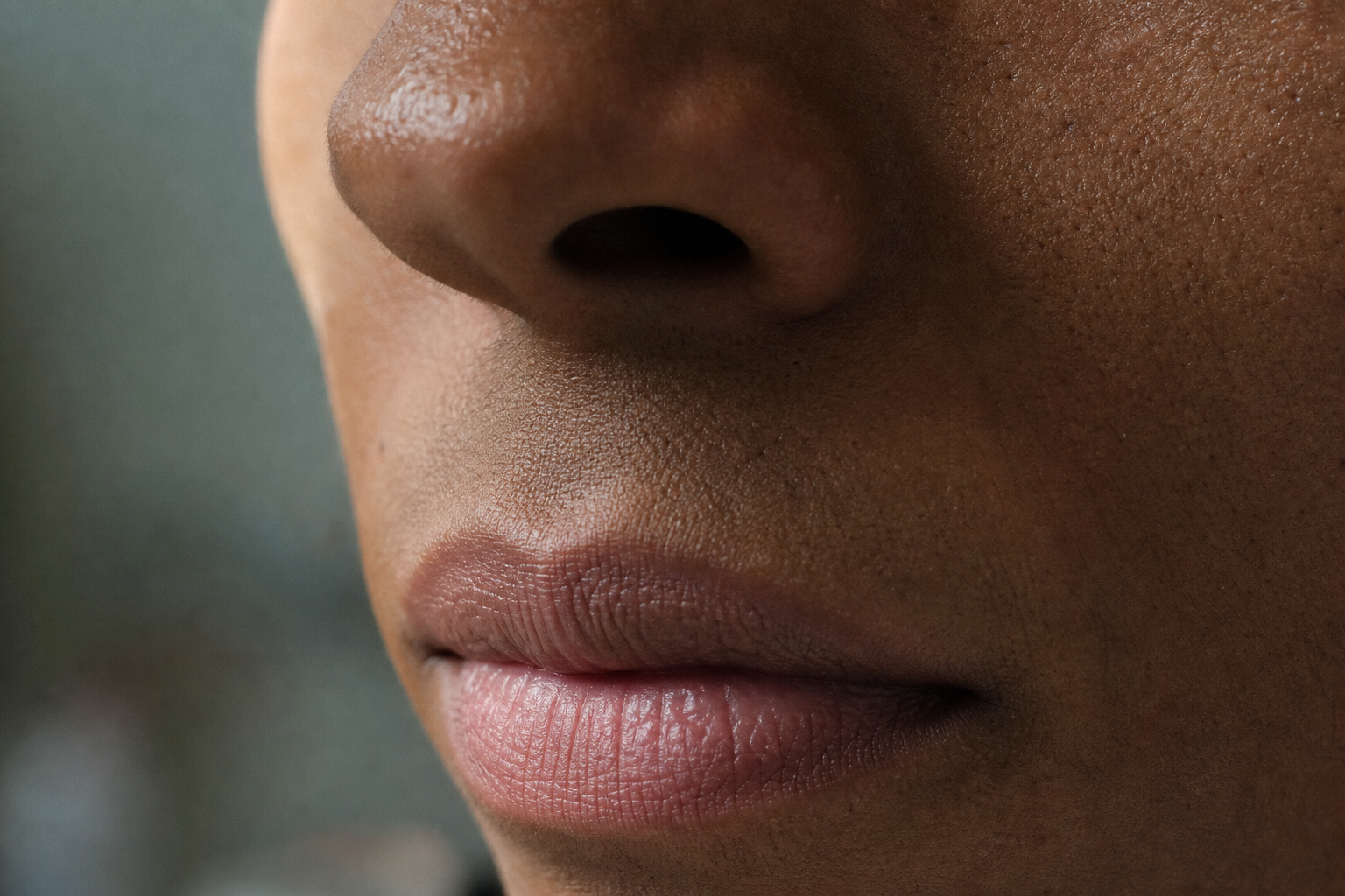
Where the pustules stop tells rosacea from perioral dermatitis.
Perioral dermatitis and papulopustular rosacea look nearly identical, but the skin right against your lip line separates them. The boundary rule, and how to photograph it for your dermatologist.
The cream that clears both, then picks a side
A dab of hydrocortisone on the bumps around your mouth, and within a few days they fade. Two weeks later they come back angrier and reach farther across your face. The steroid didn't wear off. It fed the thing it soothed. Topical corticosteroids quiet both perioral dermatitis and papulopustular rosacea in the short term, and both can rebound when you stop, but only one of them is driven by the steroid itself. Before anyone can tell you which condition is on your face, someone has to see where the pustules actually sit. That's a photography problem, and written descriptions have been losing at it for years.
Distribution is the tell
Perioral dermatitis and papulopustular rosacea read almost identically in a symptom list. Both bring small pustules around the nose and mouth. Both flush. Both sting when the wrong product lands on them. The separation lives in geography. We call it the boundary rule: perioral dermatitis spares a narrow band of skin pressed right against the lip line, a clear zone usually a few millimeters wide, and clusters just past it around the mouth, the nasal folds, and sometimes the eyelids. Papulopustular rosacea ignores that band and spreads across the central cheeks, nose, chin, and forehead. That strip of calm skin hugging the vermilion border, the pink edge of the lip, is the closest thing to a fingerprint these two conditions leave behind.
The strip of calm skin against the lip is the closest thing to a fingerprint these two conditions leave.
Why 'perioral dermatitis vs rosacea' has no satisfying answer online
Type that question into a search bar and the top results converge on the same move: a paragraph of overlapping symptoms, a shrug toward 'see a dermatologist,' and no way to act before that appointment. The reason isn't laziness. It's that the distinguishing feature is spatial, and a spatial feature dies in prose. You can't describe a two-millimeter band of sparing in a sentence and expect a worried reader to map it onto their own face in a bathroom mirror. Rosacea itself was reorganized around visible features in the 2017 update from the National Rosacea Society Expert Committee (Gallo et al. 2018), which retired the old subtype labels in favor of phenotype, meaning what you can actually see. Perioral dermatitis never got that kind of reader-facing map at all.
What the anatomy actually shows
Line the two up by footprint and they stop looking alike. Perioral dermatitis is periorificial, meaning it gathers around the openings of the face, the mouth, nose, and eyes, while conspicuously skipping the skin immediately against the lips Tempark T, Shwayder TA authored 'Perioral dermatitis: a review of the condition with special attention to treatment options,' Am J Clin Dermatol 2014;15:101–13. ([source](https://pubmed.ncbi.nlm.nih.gov/24623018/)). Rosacea is centrofacial, anchored to the convex center of the face. On lighter skin the flush does most of the pointing. On brown and Black skin it often can't: erythema reads as subtle duskiness or barely registers, which is exactly the failure mode documented when visual dermatology tools were tested across skin tones (Adamson & Smith 2018; Daneshjou et al. 2022). When color is unreliable, the arrangement of the pustules carries the whole signal, which makes the distribution map more important on darker skin, not less.
What you're mapping
Perioral dermatitis
Papulopustular rosacea
Skin right against the lip line
Spared: a clear band a few mm wide
Not spared: lesions can reach the lip edge
Where clusters gather
Mouth, nasal folds, sometimes eyelids
Central cheeks, nose, chin, forehead
Background flushing
Often minimal; fine scale and papules
Persistent central-face redness common
Clue in the history
Recent topical, inhaled, or nasal steroid use
Heat, alcohol, sun, hot drinks
General clinical patterns, not a diagnosis. Bring the photos; let your dermatologist read them. Perioral-sparing source pending verification per the note above.
The cortisone trap
The steroid history is a diagnostic clue hiding inside a treatment. Corticosteroids, whether a cream, an inhaler, or a nasal spray, are a well-recognized trigger for perioral dermatitis, and stopping them sets off a rebound flare that looks like the condition getting worse right when it should be settling Topical steroid use on the face can trigger perioral dermatitis; abrupt cessation of corticosteroids can cause rebound flaring. ([source](https://www.ncbi.nlm.nih.gov/books/NBK525968/)). That rebound loop is why the condition drags on: each flare invites another round of the cream that caused it. Rosacea can flare on steroid withdrawal too, which is why the pattern alone won't settle it. A timeline that pairs steroid use with a rebound, laid next to photos that show the spared lip band, is a far stronger story to walk into a derm office with than 'the bumps keep coming back.'
The rebound is data, not a setback
A flare worse than before after you stop a steroid cream is one of the most useful things you can document. Photograph it before reaching for anything else, and bring the sequence to your appointment. Starting, stopping, or switching any treatment is your dermatologist's call, not a search result's.
What a good perioral photo actually captures
Most phone photos of this region fail the same way: shot too far back, lit from overhead, and cropped so the lip line falls out of frame, which is the one landmark that matters. To capture the boundary at something close to clinical resolution, get near enough that the pixels resolve individual papules, hold the camera square to the skin rather than angled up or down, and keep the vermilion border inside the frame so the spared band, if there is one, is visible. Same distance, same light, same angle, every time, so week-to-week shots actually compare. This is the workflow Skinframe is built around: close-up, on-device, consistent framing, a log you can scroll to watch whether that clear band around the lips holds or fills in. The phone already in your pocket is a better distribution mapper than any symptom checklist, if it's pointed the right way and kept honest over time.
Bring the map, not the guess
You don't need to name your own condition, and you shouldn't try to. The two look alike on purpose, they overlap, and yes, you can have both at once, which is exactly why a single glance in a mirror or one clinic photo under fluorescent light can mislead. What you can do is arrive with evidence: a dated sequence of close-ups showing where the pustules sit relative to your lip line, plus a note of any steroid you've used. That turns a five-minute appointment into a reading of real data instead of a reconstruction from memory. Track the pattern, keep your own photos, and let your dermatologist make the call. That's the whole job.
Start a close-up log before your next dermatologist visit, so the distribution tells the story for you.
We build Skinframe by reading the dermatology literature first: the 2017 phenotype update, the skin-tone equity work of Adamson & Smith and Daneshjou, and the periorificial-dermatitis reviews that describe the spared lip band. The app doesn't diagnose anything. It gives you a consistent, on-device photo record precise enough for the person who does.